Frequently Asked Questions
During Maitland Grades I–II mobilizations, Type I mechanoreceptors, also known as Ruffini endings, are primarily activated, engaging specific neurophysiological pathways that modulate proprioceptive and tactile feedback. These low-threshold, slowly adapting mechanoreceptors are located in the superficial layers of joint capsules and ligaments, responding to sustained pressure and stretch. Activation of Type I mechanoreceptors leads to the transmission of afferent signals via the dorsal column-medial lemniscal pathway, which ascends through the spinal cord to the brainstem, specifically the nucleus gracilis and nucleus cuneatus. From there, the signals are relayed to the thalamus and subsequently to the somatosensory cortex, where they contribute to the perception of joint position and movement. Additionally, these mechanoreceptors play a role in modulating the gamma motor neuron activity, influencing muscle tone and reflex arcs, thereby contributing to the overall neuromodulatory effects observed during low-grade mobilizations.
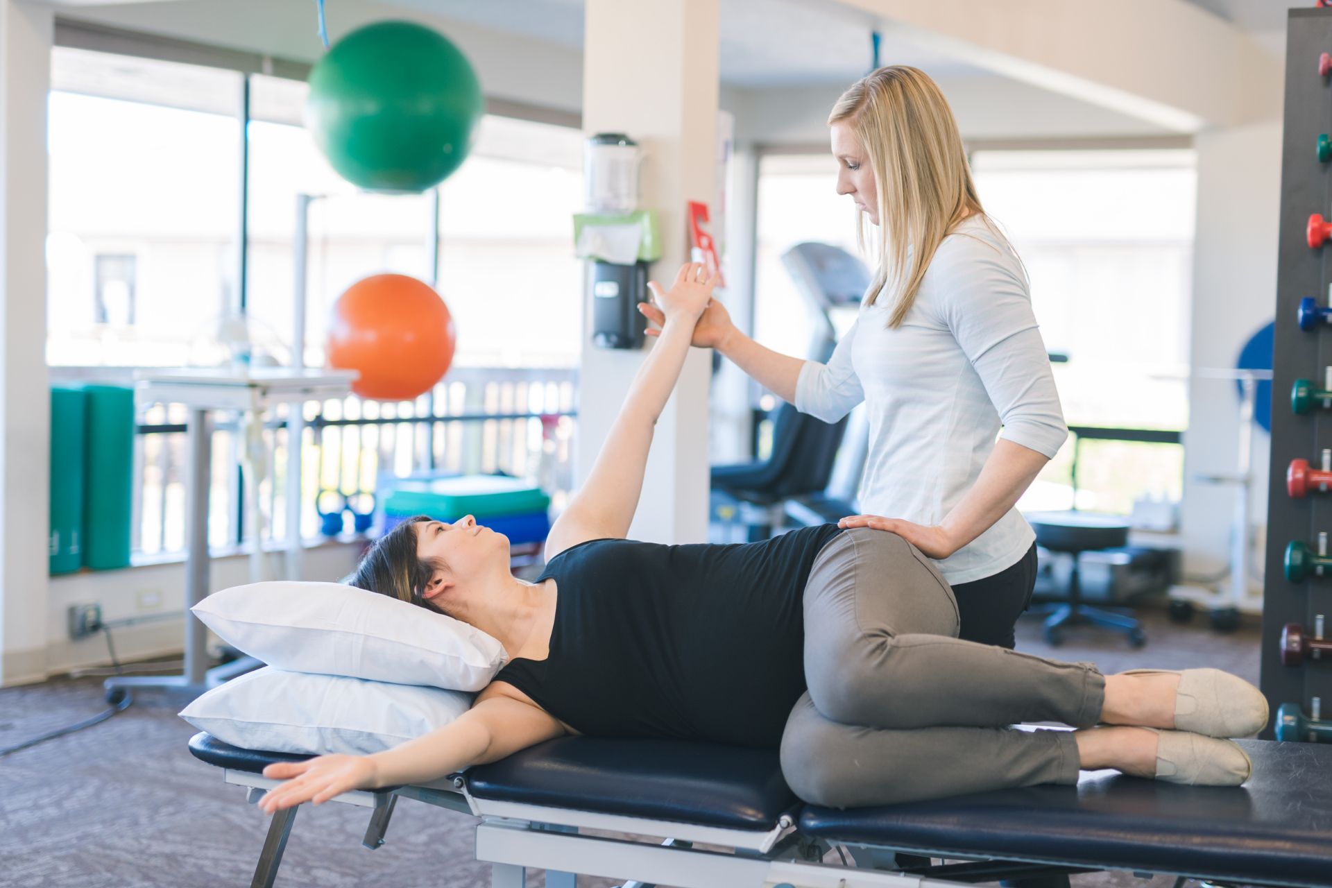
Type II mechanoreceptors play a crucial role in pain modulation during Maitland Grades I–II techniques by providing proprioceptive feedback and inhibiting nociceptive pathways. These low-threshold, rapidly adapting mechanoreceptors are primarily located in joint capsules and ligaments, responding to light touch and joint movement. When stimulated through gentle oscillatory movements characteristic of Grades I–II mobilizations, Type II mechanoreceptors activate afferent pathways that enhance the release of inhibitory neurotransmitters such as gamma-aminobutyric acid (GABA) and endorphins. This neurochemical cascade leads to the suppression of pain signals transmitted by C-fibers and A-delta fibers, thereby reducing the perception of pain. Additionally, the activation of these mechanoreceptors contributes to the gating mechanism described in the Gate Control Theory of pain, where non-nociceptive input effectively "closes the gate" to nociceptive transmission at the spinal cord level. Consequently, the gentle mobilizations in Maitland Grades I–II not only facilitate joint mobility but also promote analgesia through the strategic engagement of Type II mechanoreceptors.
Mechanoreceptor density and distribution significantly influence the effectiveness of Maitland Grades I–II mobilizations by modulating the sensory feedback and proprioceptive input to the central nervous system. These mobilizations, characterized by low-amplitude, rhythmic oscillations, primarily target superficial mechanoreceptors such as Meissner's corpuscles and Merkel's discs, which are densely populated in areas like the skin and joint capsules. The high density of these mechanoreceptors enhances the detection of subtle mechanical stimuli, facilitating the modulation of nociceptive pathways and promoting analgesic effects through the gate control theory of pain. Additionally, the distribution of mechanoreceptors, including Ruffini endings and Pacinian corpuscles, plays a crucial role in detecting changes in joint position and movement, thereby improving joint proprioception and neuromuscular control. This intricate interplay between mechanoreceptor density and distribution ensures that Maitland Grades I–II mobilizations effectively reduce pain, enhance joint mobility, and improve overall functional outcomes in patients.
The stimulation of Type I and II mechanoreceptors during Maitland Grades I–II mobilizations significantly enhances proprioceptive feedback by activating low-threshold, slowly adapting mechanoreceptors located in joint capsules and ligaments. These mechanoreceptors are sensitive to small amplitude oscillatory movements, which are characteristic of Grades I and II mobilizations. The gentle oscillations increase afferent input to the central nervous system, facilitating the modulation of neuromuscular control and joint position sense. This heightened sensory input contributes to improved kinesthetic awareness and joint stability, as the mechanoreceptors relay information about joint position, movement, and tension changes. Consequently, the activation of these mechanoreceptors aids in the reduction of pain perception through the gate control theory, enhancing the overall therapeutic effect of the mobilization technique.
Type I and Type II mechanoreceptors exhibit distinct neural responses during Maitland Grades I–II mobilizations, which are characterized by their low-amplitude, oscillatory movements. Type I mechanoreceptors, primarily located in joint capsules and ligaments, are slow-adapting and respond to static and dynamic changes in joint position, providing continuous feedback about joint position and movement. They are sensitive to low-threshold stimuli and contribute to proprioceptive awareness and postural control. In contrast, Type II mechanoreceptors, found in similar locations, are fast-adapting and respond primarily to dynamic changes, such as the onset and cessation of movement, rather than sustained pressure. They are activated by rapid, low-amplitude oscillations typical of Grades I–II mobilizations, facilitating reflexive muscle relaxation and modulation of pain through the gate control theory. The differential activation of these mechanoreceptors influences the central nervous system's processing of sensory input, affecting motor control, proprioception, and pain perception during therapeutic interventions.